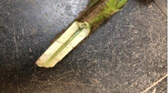
Figure 2. Interveinal chlorosis and necrosis caused by triazole phytotoxicity.

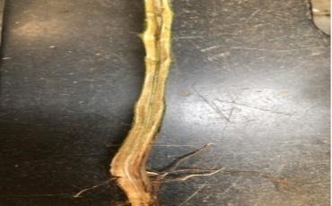
Figure 3. Lower stem and crown tissue of soybean with symptoms of triazole phytotoxicity. Note the lack of internal vascular discoloration that is commonly associated with Sudden Death Syndrome or Brown Stem Rot.
The prevalence of Group 11 (QoI a.k.a. Strobulurin) fungicide-resistant Frogeye Leaf Spot across Nebraska has led to more farmers using other fungicides and tank mixes with multiple modes of action to achieve control. One of the main groups of fungicides being utilized to control QoI-resistant frogeye leaf spot are triazoles (Group 3/DMI) fungicides
Triazoles can vary in their effects resulting in phytotoxicity and the level of injury depends on fungicide rate, adjuvants used, soybean genetics and environmental conditions at the time of application. Symptoms have been appearing 2-3 weeks after fungicide application and follow a spray pattern or have a field-wide distribution. Triazoles are xylem-mobile, so their movement through the plant is dependent on the moisture. Symptoms can be amplified when the fields are under drought stress due to the xylem-mobile nature of triazole fungicides. If the plants are drought-stressed, the fungicide remains in the leaf tissue longer, resulting in greater injury. Note that this injury will be on the leaves in the upper canopy that are expanding at the time of application.

Figure 4. Vascular discoloration of lower stem and crown commonly associated with Sudden Death Syndrome.

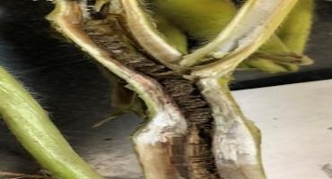
Figure 5. Internal browning and “stacking” of pith associated with Brown Stem Rot.
Sudden death syndrome (SDS) is caused by the soilborne fungus Fusarium virguliforme. Foliar symptoms of SDS typically appear at R3 and later and consist of chlorotic (yellow) spots between the veins of the upper leaves. The chlorotic spots eventually turn brown (necrotic) as the tissue dies. Leaves may drop, but the petioles remain attached. The taproot of SDS-infected plants typically rots away, otherwise below-ground symptoms of SDS are difficult to distinguish from other root rot pathogens. When splitting the stems, the vascular tissue of the lower stem/crown will be brown and discolored, but the upper portion of the stem pith remains white (Figure 4).
Brown stem rot (BSR) is caused by the soilborne fungus Cadophora gregata. Similar to SDS, this fungal pathogen causes interveinal chlorosis and necrosis of the upper leaves; however, foliar symptoms are not always present depending on the soybean variety, fungal strain and environmental conditions in the field. Splitting the stem lengthwise reveals internal browning of the vascular tissue and pith, especially at nodes and in the lower stem (Figure 5). BSR is favored by cool, wet soils and symptoms may be suppressed during periods of low soil moisture and high temperatures.
Source : unl.edu